µCT Imaging
Our µCT imaging services support high-resolution three-dimensional imaging, quantitative structural analysis, and longitudinal in vivo and ex vivo studies using the Quantum GX2 microCT Imaging System. Combining non-destructive X-ray imaging with advanced reconstruction and morphometric analysis, the platform enables detailed visualization and characterization of bone architecture, fracture healing, soft tissue contrast-enhanced structures, implants, and biomaterials for preclinical and translational research applications.
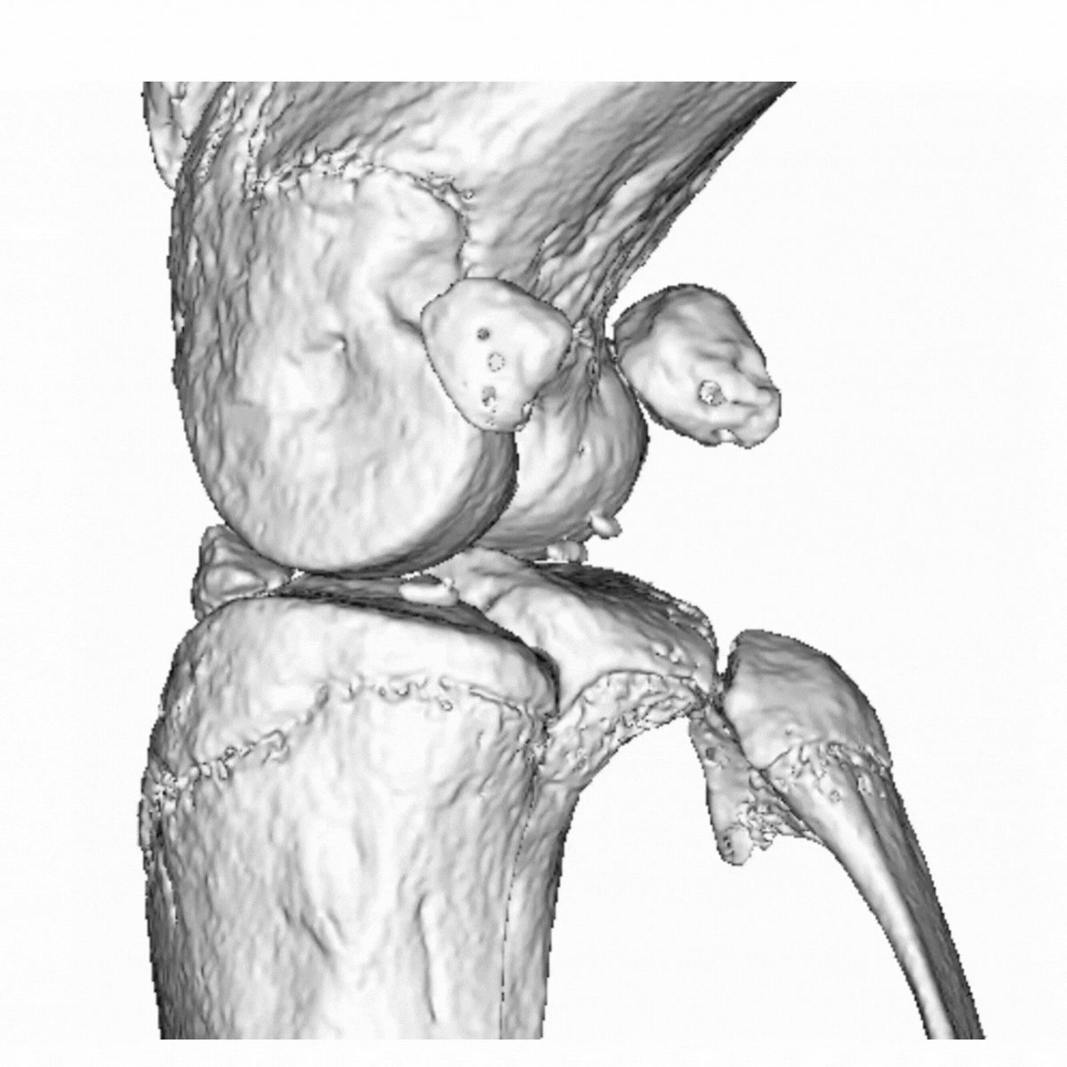
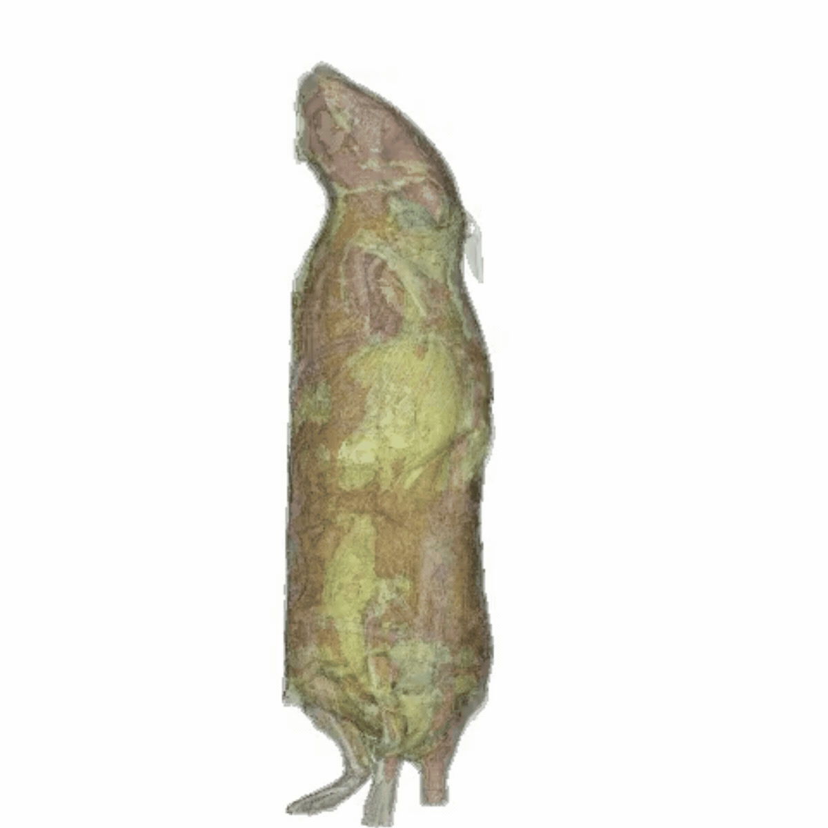
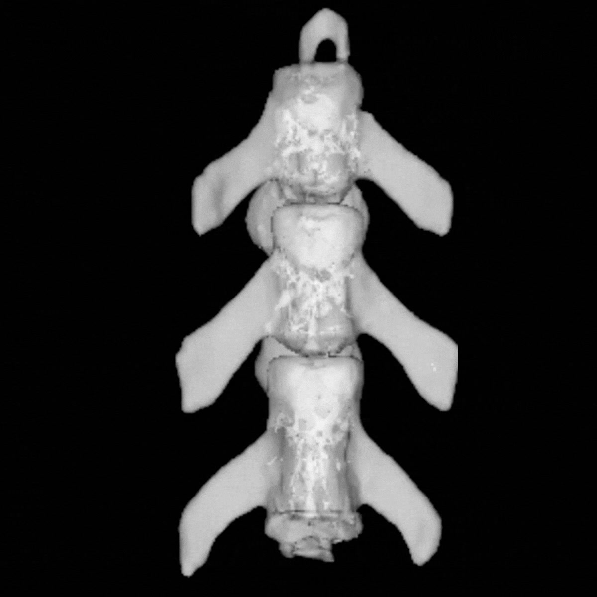
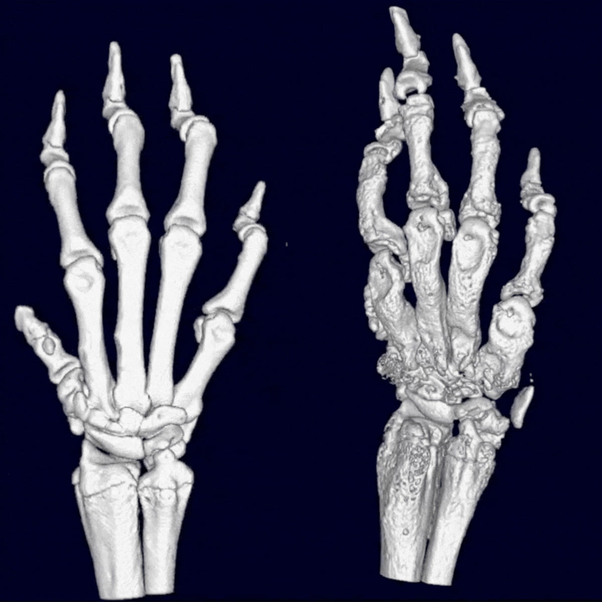
The Quantum GX2 microCT platform delivers high-resolution 3D reconstructions for morphometric analysis, longitudinal studies, and structural characterization. The animated scans below show representative reconstructions acquired with the system.
Applications
| Application | Description |
|---|---|
| Bone Morphometry | Quantitative analysis of trabecular and cortical bone architecture including BV/TV, trabecular thickness, separation, cortical thickness, and bone mineral density. |
| Fracture and Bone Healing Models | Monitoring of callus formation, bone regeneration, fixation stability, and healing progression in longitudinal studies. |
| Arthritis Models | Evaluation of bone erosion, osteophyte formation, joint destruction, and paw inflammation in models such as CIA. |
| Osteoporosis Research | Assessment of bone loss, microarchitectural deterioration, and treatment efficacy in preclinical osteoporosis models. |
| Orthopedic Implant Analysis | Visualization and evaluation of implant positioning, osseointegration, and surrounding bone remodeling. |
| Whole Body Imaging | Non-invasive imaging of complete small animals for skeletal screening, anatomical visualization, and body composition studies. |
| Soft Tissue Contrast Imaging | Imaging of vascular structures or soft tissues using contrast agents for enhanced visualization. |
| Longitudinal in vivo Studies | Repeated imaging of the same animal over time to monitor disease progression or therapeutic response while reducing animal numbers. |
| ex vivo Tissue Analysis | High-resolution imaging of isolated organs, bones, or biomaterials with maximal image quality and extended scan settings. |
| Biomaterial and Scaffold Characterization | Structural analysis of biomaterials, porous scaffolds, and tissue-engineered constructs. |
Key Features
- High-resolution 3D imaging for in vivo and ex vivo applications
- Non-destructive analysis allowing downstream histology and molecular analyses
- Quantitative structural and morphometric analysis
- Longitudinal imaging capabilities for therapy monitoring
- Fast acquisition with flexible scan settings
- Compatible with bone, soft tissue, implant, and biomaterial studies
- Advanced 3D reconstruction and visualization workflows
- Reduction of animal numbers through repeated measurements in the same subject