Technology
Confocal Imaging
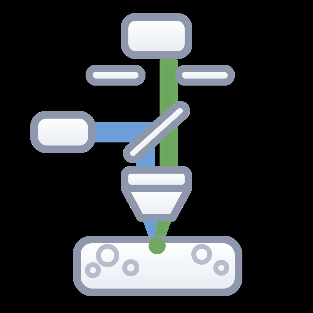
Our confocal imaging services support medium-throughput cellular imaging, phenotypic characterization, and longitudinal biological studies using the Agilent BioTek Cytation C10 Confocal Imaging Reader. Combining laser-based confocal imaging with automated image acquisition and quantitative analysis, the platform enables high-quality fluorescence and brightfield imaging for preclinical, translational, and cell biology research in multiwell plate formats.
Applications
| Application | Description |
|---|---|
| Phenotypic Screening | Quantitative assessment of cellular morphology, viability, proliferation, and functional responses. |
| Live-Cell Imaging | Time-lapse monitoring of cellular behavior, migration, growth, and treatment responses. |
| Immunofluorescence Analysis | Visualization and quantification of protein expression and intracellular localization. |
| 3D Cell Model Analysis | Phenotypic analysis of engineered and primary 3D cell populations, including spheroids and organoids. |
| Cell Therapy Research | Characterization of engineered and primary cell populations using multiplex fluorescence imaging. |
| Longitudinal Studies | Standardized image acquisition and analysis across multiple study time points. |
| Inflammation & Toxicity Research | Analysis of cellular activation, structural integrity, and inflammatory or cytotoxic responses. |
| Translational Studies | Integration of imaging endpoints into efficacy, biodistribution, and mechanism-of-action workflows. |
Key Features
- LDI-7 Laser Illuminator providing stable, high-intensity excitation for multiplex fluorescence imaging
- Laser-scanning confocal imaging for improved optical sectioning and background reduction
- Laser autofocus system ensuring accurate focus maintenance during automated imaging workflows
- Multi-mode imaging capabilities including: Confocal fluorescence imaging; Widefield fluorescence imaging; Brightfield imaging; Phase contrast imaging.
- Compatible with standard multiwell plate formats for medium-throughput screening applications
- Automated image acquisition and analysis workflows
- Quantitative single-cell and population-level measurements
- High-resolution imaging with a range of objectives optimized for screening and detailed cellular analysis
- Reproducible longitudinal image acquisition across complex study designs
- Flexible assay development and image analysis support